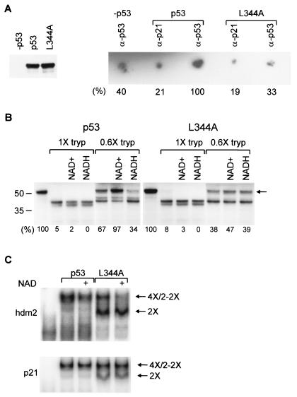
FIG. 3.
NAD+ binding, affecting p53 conformation and specifically inhibiting p53 tetramer DNA binding in vitro. (A, left) Equal amounts of p53 and L344A were incubated with 32P-NAD+. (Right) Bound 32P-NAD+. Signal strength is indicated below each spot; signal strength for anti-p53 is arbitrarily 100%. (B) [35S]methionine-labeled proteins were incubated with NAD+ or NADH and then digested with trypsin (tryp). The percentage of undigested p53 (arrow) is indicated below each lane. (C) NAD+ was added with 32P-hdm2 (top) or 32P-p21 (bottom) to p53 tetramers or dimers. DNA-bound proteins are tetramers (4×), single L344A dimers (2×), or pairs of L344A dimers (2-2×).

