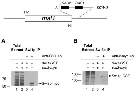
FIG. 5.
Interaction of Swi1p with Swi3p in the smt-0 mutant. A schematic representation of the 263-bp deletion of the smt-0 mutation is shown at the top. The deletion includes both SAS1 and SAS2 sequences, shown as black boxes. Immunoprecipitation (IP) was performed as described for Fig. 3 and 4. (A) Total protein extracts from SP982 (swi1 swi3) and BSP50 (swi1-GST swi3-myc) were loaded in lanes 1 and 2, respectively. With the total protein extract of BSP50, a mock precipitate of Swi1p-GST with protein A-agarose beads (lane 3) and the immunoprecipitate of the protein with anti-GST antibody (lane 4) were analyzed for Swi3p-myc by Western analysis. (B) Total protein extracts from SP982 and BSP50 were loaded in lanes 1 and 2, respectively. A mock precipitate of Swi3p-myc with protein G-agarose beads and the immunoprecipitate of Swi3p-myc with anti-c-myc antibody were loaded in lanes 3 and 4, respectively. The gel was subjected to Western analysis to detect Swi1p-GST.

