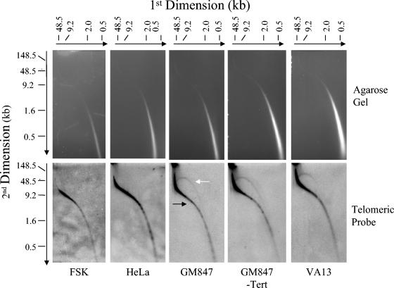
FIG. 4.
2D PFGE of TRFs from ALT and non-ALT cells. Total DNA (20 μg) was digested with HinfI/HaeIII and then separated by 2D PFGE. Telomeric material was detected by in-gel hybridization with a [γ-32P](CCCTAA)6 probe and was visualized using a PhosphorImager. The black and white arrows indicate linear- and circular-form DNA, respectively.

