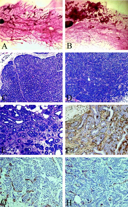
FIG. 3.
Whole-mount analysis and histological characteristics of WT (A, C, G) and IRS-2 null (B, D-F, H) PyV-MT mammary tumors. (A and B) The number 4 inguinal mammary glands from WT and IRS-2−/−, PyV-MT+/− mice were fixed and stained with carmine alum stain. Bar, 1 mm. (C and D) Solid nodular high-grade tumors that lack glandular structure and contain minimal stroma. (E) Glandular, low-grade tumor region from an IRS-2−/− mouse. Myoepithelial cells are still present surrounding the dilated glands, as demonstrated by the positive smooth-muscle actin staining (F). (C-F) Bar, 100 μm. (G and H) Mammary tumors from WT (G) and IRS-2−/− (H) mice stained with CD31 to detect tumor angiogenesis. Bar, 50 μm.

