
Figure 2. Partitioning of the prostate for the user study.
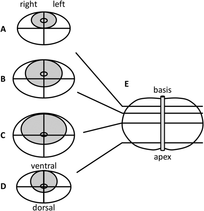
Here, it is shown how the prostate was divided into segments for radiological and histopathological evaluation. (A–D) the four axial cuts that were considered, (E) sagittal view of the prostate and urethra.