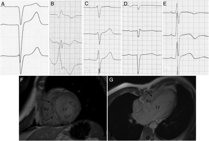
Figure 3.
(A–E) Example ECGs presenting septal remodelling. (A) Q waves in V1–V2; (B) broad QRS with fragmentation in V2–V3, and a Q wave in V1; (C) RV1>RV2 with fragmented QRS in V2; (D) poor R-wave progression with QRS fragmentation in V2; (E) RV2>RV3 with fragmented QRS in V1. 3. (F and G) CMR image of an LMNA mutation carrier. Arrows point to the areas showing late gadolinium enhancement. CMR, cardiac MRI; LV, left ventricle; RV, right ventricle.

