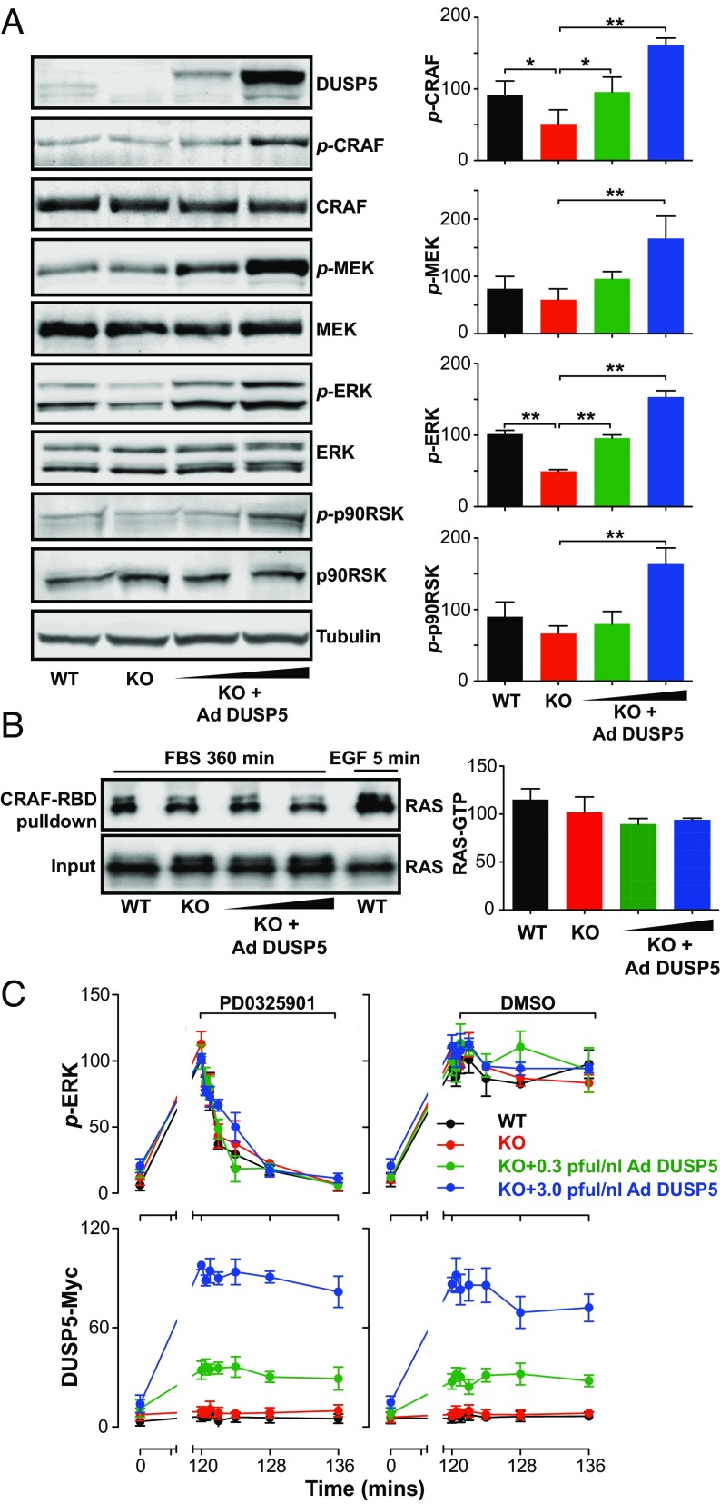
Fig. 2.
DUSP5 propagates ERK signaling by increasing RAF and MEK activity. WT and Dusp5 KO primary MEFs were infected with 0.3–3.0 pfu/nL Ad EGR1 promoter-driven DUSP5-Myc (Ad DUSP5) as indicated. (A) MEFs were stimulated for 360 min with 20% (vol/vol) FBS. Western blots of whole-cell lysates were probed for p-sites on ERK (TEY activation loop), MEK (Ser217/221), CRAF (Ser338), and p90RSK (Thr-359/Ser363) as well as total kinase levels and β-tubulin. Normalized blot quantification is shown ± SEM, n = 4. *P < 0.05, **P < 0.01 comparing KO vs. all columns using one-way ANOVA and Dunnett's posttest. (B) MEFs were stimulated either with 20% (vol/vol) FBS for 360 min or 50 ng/mL EGF for 5 min before lysis and pull-down of GTP-RAS using the RAS-binding domain (RBD) of CRAF. Input levels of total RAS in whole-cell lysates and levels of GTP-RAS in the pull-down were measured by Western blotting. Normalized mean blot quantification of GTP-RAS is shown ± SEM, n = 3. (C) MEFs were stimulated with 20% (vol/vol) FBS for 120 min before addition of either DMSO vehicle or 5 μM PD0325901 MEK inhibitor for times indicated. Levels of Myc-tag and whole-cell levels of p-ERK were measured using HCM. Data shown are normalized population AFU values, n = 3 ± SEM.

