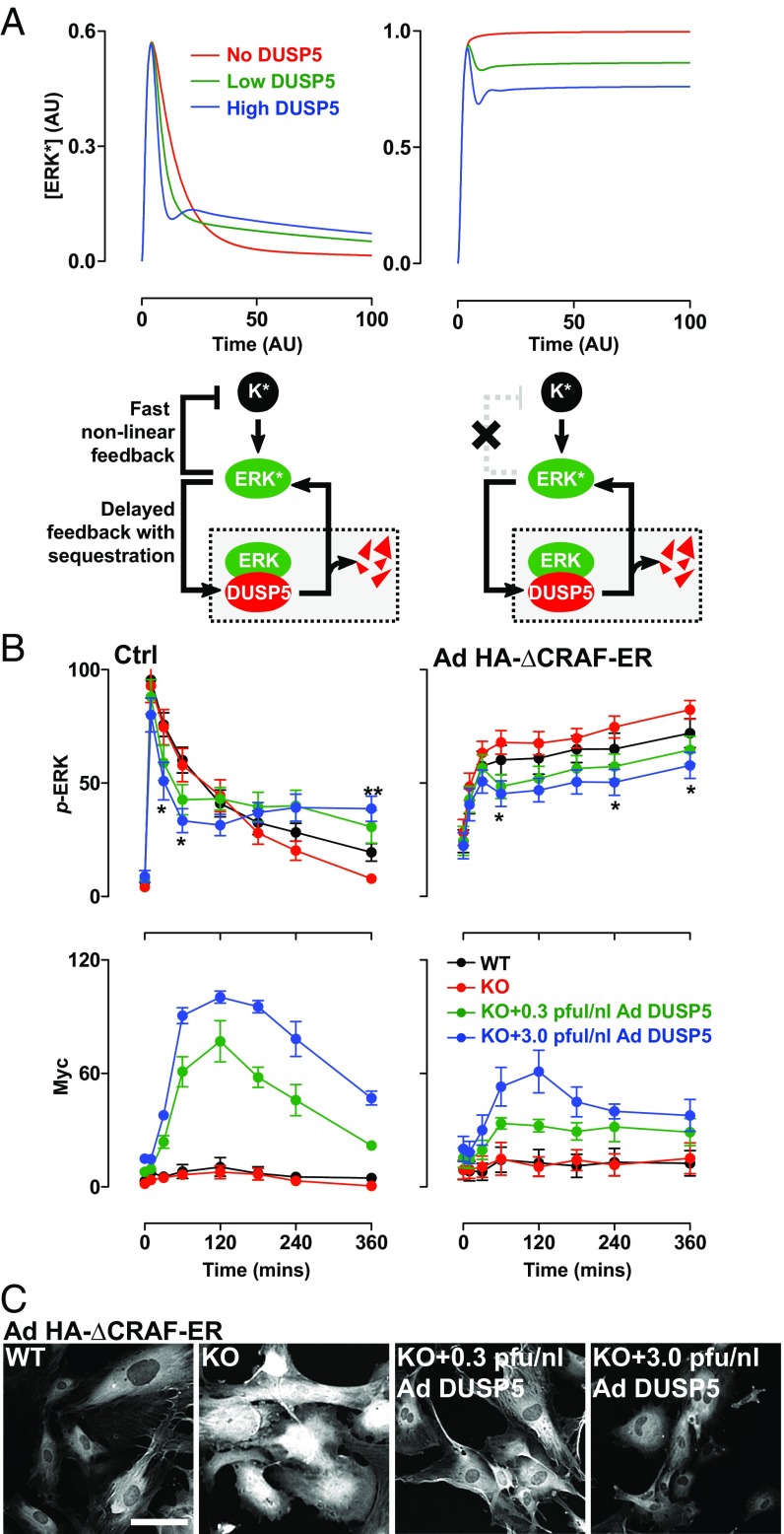
Fig. 4.
DUSP5 propagates ERK signaling by relieving upstream kinase inhibition. (A) Conceptual model network structures comprising ERK activation by an upstream kinase (K), feedback inhibition and sequestration of ERK by DUSP5, and nonlinear feedback inhibition of K by ERK. K* and ERK* denote activated forms of K and ERK, respectively. The plots show model predictions of ERK* concentration vs. time in arbitrary units (AU) under conditions where there is no, low, or high levels of DUSP5 synthesis and in which negative feedback between ERK and K is intact (Left) or completely disabled (Right). (B) WT and Dusp5 KO MEFs were infected with either empty Ad, or Ad expressing HA-ΔCRAF-ER, alongside either 0.3 or 3 pfu/nL of Ad EGR1 promoter-driven DUSP5-Myc. Cells were stimulated with either 20% (vol/vol) FBS (Left) or 0.1 µM 4HT (Right) before immunostaining for p-ERK and Myc followed by HCM analysis. Normalized population mean AFU values for whole-cell p-ERK and Myc intensity are shown ± SEM, n = 4–7, *P < 0.05, **P < 0.01 comparing KO vs. KO + 3.0 pfu/nL Ad DUSP5-Myc using two-way repeated-measures ANOVA and Bonferroni posttest. (C) Representative HCM images of p-ERK staining are shown from experiments described in B, comparing Ad HA-ΔCRAF-ER–infected MEFs after 360-min stimulus with 0.1 µM 4HT. (Scale bar: 100 μm.)

