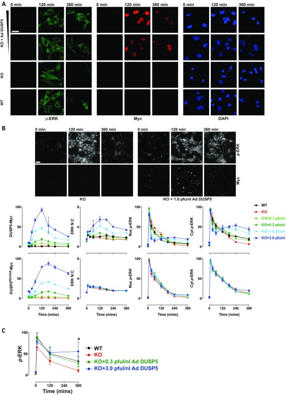
Fig. S1.
DUSP5 increases cytoplasmic ERK responses. Primary WT or Dusp5 KO MEFs were infected with either empty Ad or 0.1–3.0 pfu/nL Ad ERK-dependent EGR1 promoter-driven DUSP5-Myc or a KIM mutant (DUSP5R53/54A-Myc) before stimulation with 20% (vol/vol) FBS as indicated. (A) Representative images are shown for p-ERK, Myc-tag, and DAPI staining from confocal microscopy experiments comparing rescue using 0.3 and 3 pfu/nL Ad DUSP5. (Scale bar: 60 μm.) (B) Representative images and quantification in plots are shown for a single HCM experiment comparing the full range of DUSP5 or DUSP5R53/54A rescue conditions from 0.1–3.0 pfu/nL Ad concentrations. Note: KO data are identical in Upper and Lower plots. Data are shown as normalized population averages of AFU ± SD, n = 2–4. (Scale bar: 100 μm.) (C) Primary WT or Dusp5 KO MEFs were infected with either empty Ad or 0.3–3.0 pfu/nL Ad DUSP5-Myc before 20% (vol/vol) FBS stimulus and Western blotting as described in Fig. 1C. Data shown are normalized p-ERK levels measured in whole-cell lysates ± SEM, n = 3, *P < 0.05 comparing KO vs. KO + 3.0 pfu/nL DUSP5 using two-way repeated-measures ANOVA and Bonferroni posttest.

