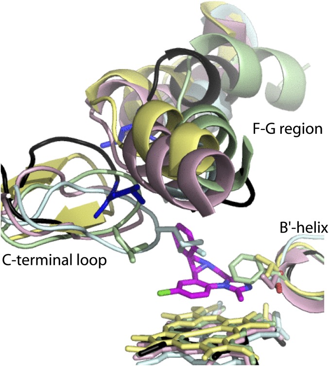
Fig. S5.
Structural overlay of human drug-metabolizing CYPs: CYP3A4 (1WOE; black), CYP1A2 (2HI4; cyan), CYP2B6 (molecule A of 4RQL; pink), CYP2C9 (molecule A of 1OG2; yellow), and CYP2D6 (molecule A of 4WNU; green). MDZ (in magenta) is shown in the crystallographic binding mode. Neither of the B′–C loop residues aligning with Ser119 of CYP3A4 can H-bond and accommodate MDZ in a similar mode. The length and topology of the F–G and C-terminal loops significantly varies among CYPs, due to which it is not possible to accurately pinpoint residues corresponding to MDZ-flanking leucines 216 and 482. Nevertheless, the outlined sequence and structural dissimilarities, and preservation of all key elements in CYP3A5 and CYP3A7 (Fig. S6) could explain why MDZ is predominantly oxidized by the CYP3A enzyme subfamily.

