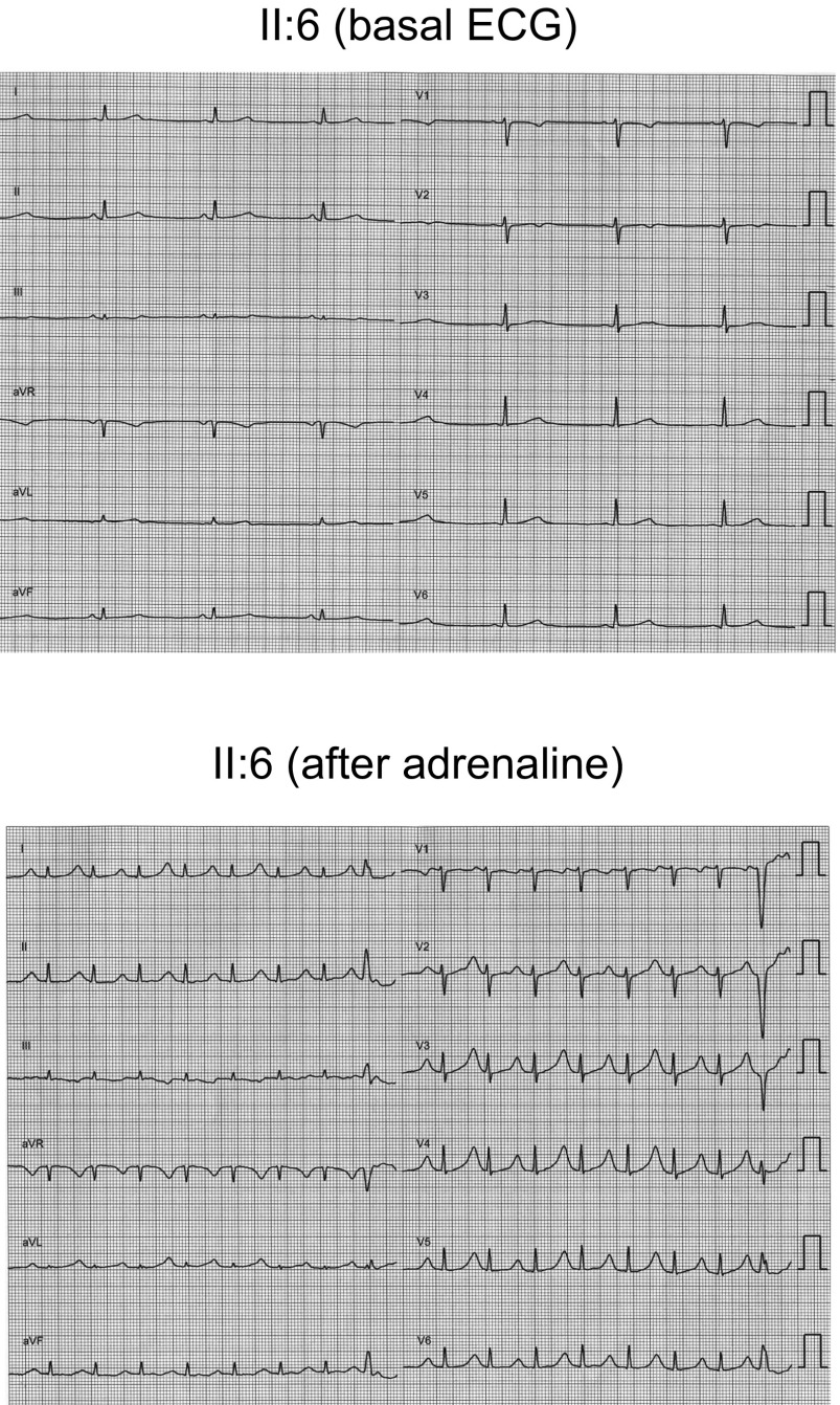
Fig. S1.
Twelve lead ECGs of sister II:6 under basal conditions (Top) and after adrenaline bolus injection (Bottom) (paper speed 25 mm/s). Basal ECG shows normal PR (140 ms), QRS (90 ms), sinus bradycardia (48 bpm), and low-voltage wide QT waves (QTc 460 ms). As shown, the adrenaline test was positive, because QTc was prolonged to 618 ms, the amplitude of the T waves exhibited alternance, and polymorphic ventricular extrasystoles were developed. Adrenaline tests were developed in all relatives following the protocol described by Shimizu et al. (33).

