Fig. 2.
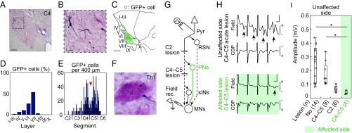
Visualization of blocked PNs and confirmation of blockade of synaptic transmission through PNs. A–F were obtained from monkey U, and G–I were obtained from monkey K. (A) Representative GFP-labeled cells in C4. Dashed square indicates area shown in B. (Scale bar, 200 μm.) (B) High magnification of A. (Scale bar, 50 μm.) (C) Reconstructed distribution of GFP-labeled cells by multiple sections, which were separated by 200 μm. I–X, laminae of Rexed. (Scale bar, 500 μm.) (D) Distribution of GFP-labeled cells in individual laminae (I–X). (E) Longitudinal distribution of GFP-labeled cells along the spinal cord. Red arrow indicates the site of lesion. (F) A representative axon and bouton of GFP-labeled cell in a motor nucleus at Th1. The sections were counterstained with Neutral Red. (Scale bar, 10 μm.) (G) The arrangement of terminal acute electrophysiological experiments. In addition to the direct cortico-motoneuronal connection, indirect pathways through reticulospinal neurons (RSN), propriospinal neurons (PNs), and segmental interneurons (sINs) might exist. During the experiments, the lateral CST was transected at C4–C5 and successively at C2. MNs, motoneurons. Pyr, contralateral medullary pyramid. (H) Representative field potentials (Field) and cord dorsum potentials (CDP) in the unaffected side of a monkey with a C4–C5 acute lesion (Top) and in the affected side (Bottom) following four trained stimuli of Pyr at 200 μA. Arrows indicate disynaptic field potentials. (Vertical scale bar, 0.2 mV; horizontal bar, 1 ms.) (I) Quantitative analysis of the amplitudes of the disynaptic field potentials with no lesion, a C4–C5 lesion, and a C2 lesion on the unaffected side, and with a C4–C5 lesion on the affected side. The numbers of records are shown in parentheses. The box plots represent the 25th and 75th percentiles of the data. *P < 0.01 (the Wilcoxon test).

