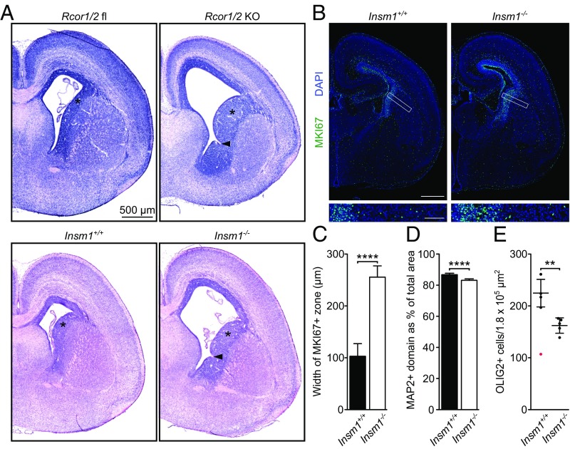
Fig. 6.
Insm1−/− mice phenocopy several Rcor1/2 knockout phenotypes in E18.5 brain. (A) H&E-stained E18.5 coronal hemisections. Asterisks indicate the VZ/SVZ; arrowheads indicate interganglionic sulci. (B) Representative hemisections immunolabeled for the proliferation marker MKI67 and counterstained with DAPI. Boxes indicate Insets shown below at higher magnification. [Scale bar, 500 μm (Top) and 100 μm (Bottom).] (C–E) Immunohistochemical analysis of coronal hemisections of Insm1+/+ and Insm1−/− forebrain. Statistical significance was assessed by t tests. The means and SDs are indicated. (C) Quantification of the width of the MKI67+ zone. Measurements were made from areas comparable to those depicted in the Insets in B. (D) Quantification of MAP2 immunolabeling, showing the percentage of each E18.5 coronal hemisection occupied by the MAP2+ domain. (E) Quantification of the number of OLIG2+ cells in the IZ of the neocortex. For each mouse, one region of 3 × 104 μm2 was selected from each of six hemisections, and the numbers of OLIG2+ cells from all regions were added. Each mouse is represented by one dot. Exploratory data analysis identified one wild-type count (107, indicated in red) as an outlier, so it was omitted from the statistical analysis. **P < 0.01; ****P < 0.0001.

