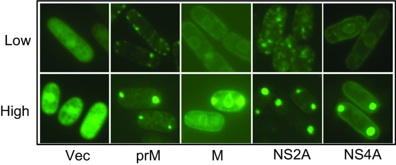
Fig. S2.
Comparison of intracellular localization patterns of ZIKV proteins under high and low levels of protein expression. Only ZIKV proteins that showed differences in localization patterns and possible cytoplasmic puncta are shown here. All GFP-ZIKV proteins were expressed from the pYZ3N gene-expression vector through the inducible nmt1 promoter. The high level of ZIKV protein was produced with full induction of the nmt1 promoter by complete removal of thiamine from the growth medium. The subcellular localizations of each ZIKV protein were observed between 24 and 48 h after GI. The low level of ZIKV protein was produced with partial induction of the nmt1 promoter using 10 nM thiamine in the growth medium (14, 51). The subcellular localizations of each ZIKV protein were observed within 20 h after GI.

