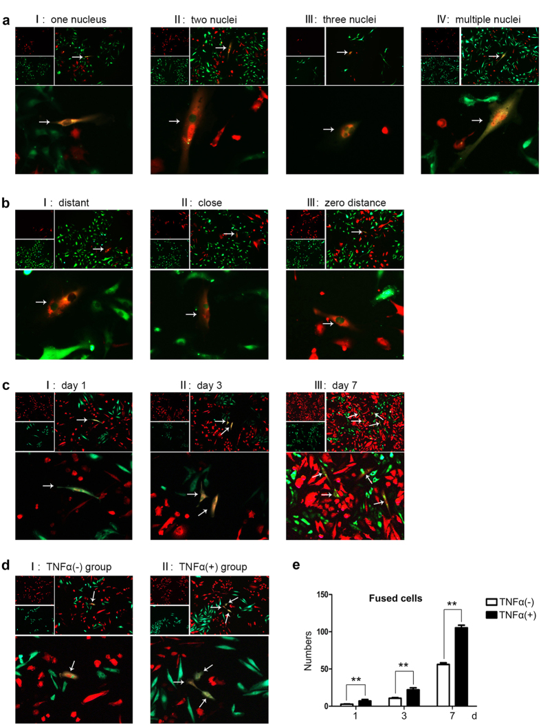
Figure 1. Spontaneous fusion between SCC-9 and HUVEC was enhanced under the stimulation of TNF-α.
(a) I–IV Representative fluorescence images show the number of nucleus in fusion cells. The number of nucleus is 1, 2, 3 and multiple in sequence from (a I–a IV). (b I–III) Representative images of fluorescence displayed the distance between nuclei in different hybrid cells. (b I) showed that the distance of nuclei in fused cells was relatively far; (b II) showed adjacent; (b III) showed the nuclei fused to one and became zero distance. (c) Condition of fused cells after co-cultured for 1, 3 and 7 days. These fused cells appeared healthy. (d) Representative images of fluorescence of fused cells between TNF-α group (d II) and normal group (d I). The stimulation of TNF-α increased the percentage of bi-fluorescent cells to 1.78 ± 0.053%, compared with untreated cells (0.748 ± 0.024%). The magnification of images a,b,c and d was 100× and 200×, respectively. (e) Number of fused cells of TNF-α group and normal group by artificial counting method. The error bars correspond to mean ± SD of at least three independent experiments. *P < 0.05, **P < 0.01, ***P < 0.001.

