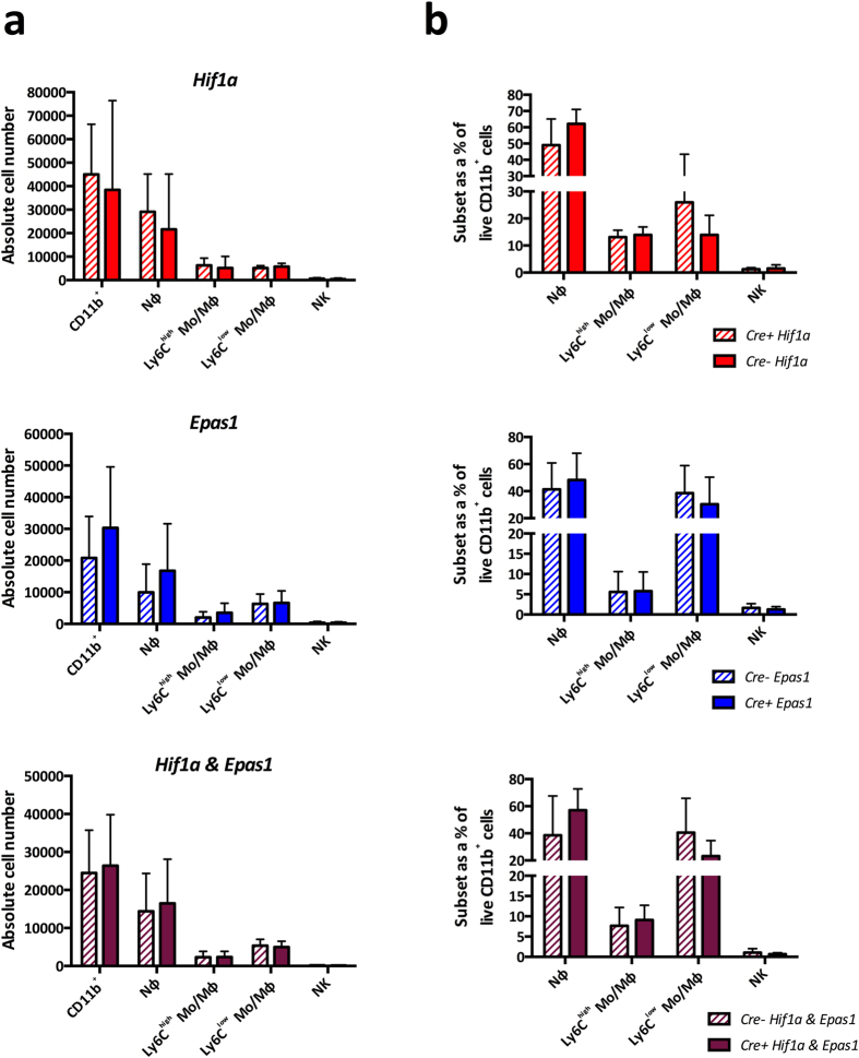
Figure 2. Deletion of HIF genes in myeloid cells does not influence EIU at peak disease.
Flow cytometric analyses of (A) absolute cell numbers and (B) proportions of myeloid subsets infiltrated in the eye 18 hours after EIU induction in animals with myeloid cells deficient in Hif1a, Epas1 and Hif1a & Epas1 with their floxed littermate controls. Myeloid cell populations are defined using standard gating strategy. Nϕ = neutrophils; Mo/Mϕ-monocyte/macrophages. Graphs show mean ± SD; n = 10–12 injected eyes per group; > 3 independent experiments.

