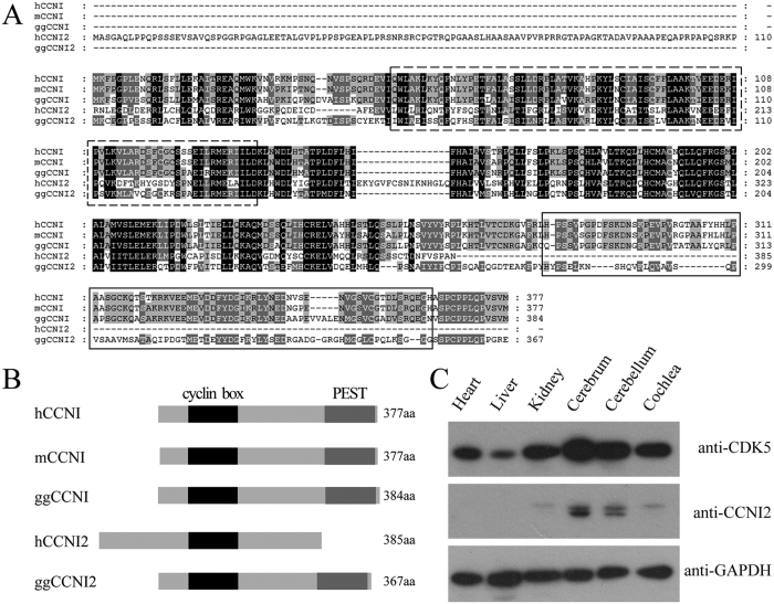
Figure 1. Protein sequence and tissue expression pattern of CCNI2.
(A) Amino acid sequence alignment of CCNI and CCNI2 from different species. Amino acid sequences of Homo sapiens CCNI, Mus musculus CCNI, Gallus gallus CCNI and Homo sapiens CCNI2, Gallus gallus CCNI2 were aligned using the ClustalW method. Dashed box indicates the cyclin box. Solid box indicates the PEST region. (B) Schematic representation of the domain structures of CCNI and CCNI2. Cyclin box and PEST region are indicated. (C) Tissue expression pattern of chicken CCNI2 was examined by western blot. Total proteins from E18.5 chicken tissues were extracted and separated by PAGE and detected with antibodies against CDK5, CCNI2, and GAPDH.

