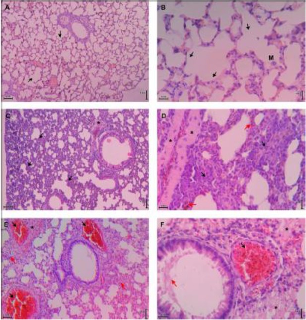
Fig. 3:
Histopathology of lung tissue of control, renal malaria and cerebral malaria groups. Images (A) and (B) are representatives of control mice lung tissue that show normal histology; no thickening in septal alveolar; black arrow indicated the normal alveoli; M=alveolar macrophage. Images (C) and (D) are representatives of lung tissue of renal malaria mice that show alveolar septal thickening 2–4 times than control due to infiltration of inflammatory cells (black arrow), vascular edema (*), and interstitial microhemorrhage (red arrow). Images (E) and (F) are representatives of lung tissue of cerebral malaria mice that show alveolar septal thickening more than 4 times compare to control (red arrow in E), exudates (*), thrombus with sequestration of iRBCs and hemozoin (black arrow), and hyaline membrane (red arrow in F). A, C, E = total magnification of 100×; B, D, F = total magnification of 400×. All tissues were stained with HE

