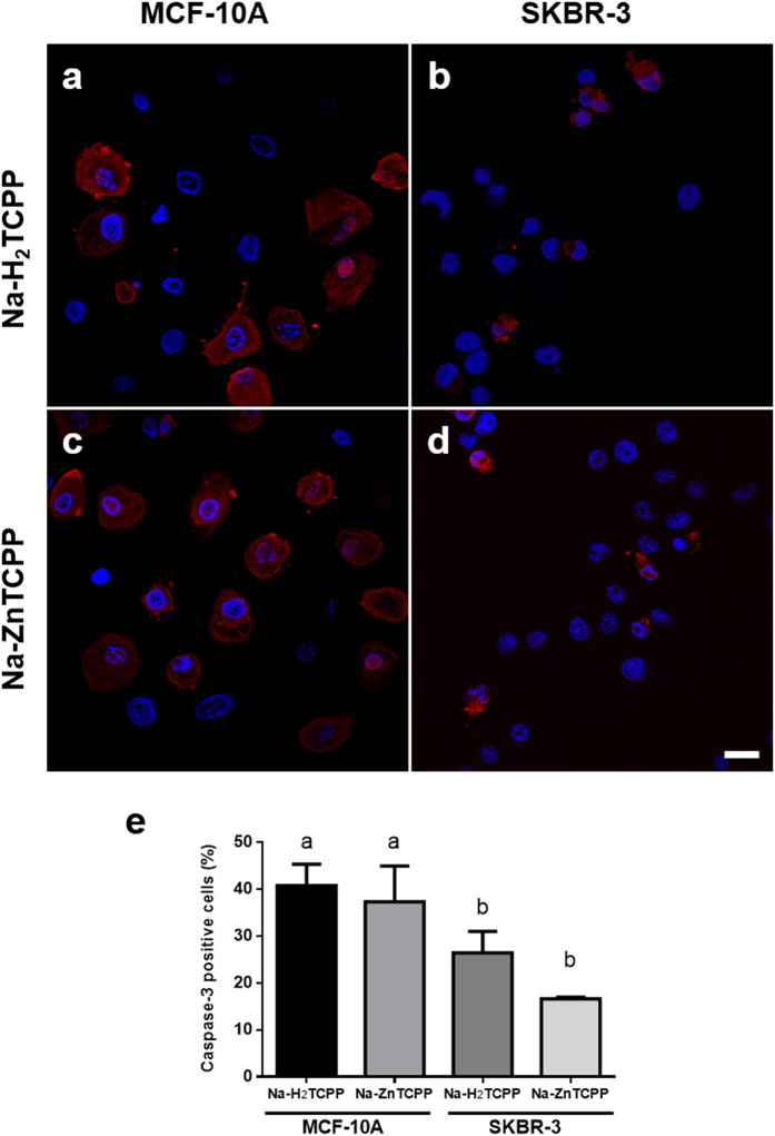
Figure 6. Cells processed for immunofluorescence staining of active caspase-3 (red) and counterstained with Hoescht-33258 (blue).
(a) MCF-10A cells incubated with 4 μM Na-H2TCPP and processed 5 h after irradiation. (b) SKBR-3 cells treated with 4 μM Na-H2TCPP and processed 5 h after treatment. (c) MCF-10A cells incubated with 4 μM Na-ZnTCPP and processed 5 h after photodynamic treatment. (d) SKBR-3 cells treated with 4 μM Na-ZnTCPP and processed 5 h after treatment. Scale bar, 20 μm. (e) Percentage of active caspase-3 positive cells observed 5 h after photodynamic treatments. Different superscripts on top of the columns denote significant differences between groups not sharing the same superscript.

