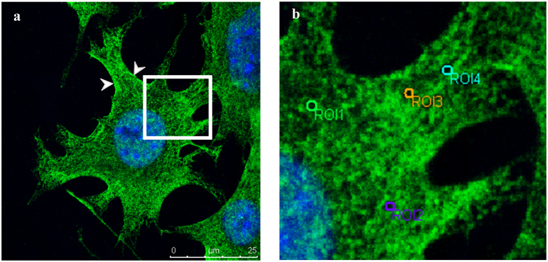
Figure 1. The spectrin of MLO-Y4 was fluorescence stained with Alexa 488 (green) and the nucleus was labeled by DAPI (blue) to locate cells.
The spectrin was observed on the cell membrane, and also in the cytoplasm and nucleus. Some intense stainings were found on the membrane (indicate by arrow heads) (a). The box inset in (a) was amplified (b), and it showed that the spectrin was organized to a porous network structure. The grid of the spectrin network was selected as a region of interest (ROI) for measurements.

