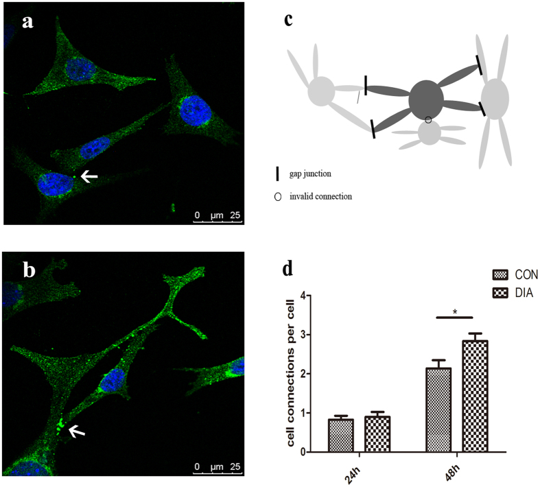
Figure 8. The Cx43 of osteocytes was fluorescent stained and the cell-cell connections of each osteocyte were counted.
(a) The Cx43 of osteocytes was fluorescent stained (green). In CON group, the Cx43 was distributed both in the cytoplasm and on the cell membrane. Sparse intense staining can be observed at cell-cell contacts. (b) After the treatment of DIA, more Cx43 can be found at cell-cell contacts at 48 h after planting. As depicted in (c), the connections between cell processes, and the connections between the cell process and cell body were counted, while the connections between the cell bodies were not counted. The cell-cell connections were not fully developed at 24 h after cell planting. At 48 h, the cell-cell connections were formed, and the treatment of DIA promoted the formation of the cell connections by 33% compare with CON (d). *p < 0.05.

