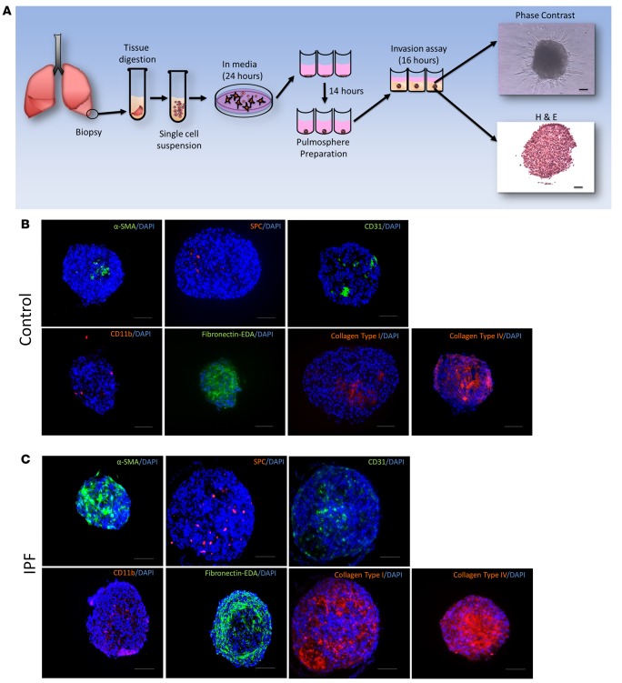
Figure 1. Pulmospheres are 3D structures made of multiple cell types and extracellular matrix.
(A) Schematic representation of pulmosphere preparation. Representative phase-contrast microscopic image. H&E-stained pulmosphere. Scale bar: 250 μm. (B) Immunofluorescent staining of pulmospheres from IPF patients for α-SMA, SPC, CD31, CD11b, fibronectin-EDA, collagen type I, and collagen type IV in paraffin-embedded sections. All immunofluorescent slides were costained with DAPI for nuclei. Five random pulmospheres were sampled from each control subject (n = 9). Scale bar: 250 μm. (C) Immunofluorescent staining of pulmospheres from IPF patients for α-SMA, SPC, CD31, CD11b, fibronectin-EDA, collagen type I, and collagen type IV in paraffin-embedded sections. All immunofluorescent slides were costained with DAPI for nuclei. Five random pulmospheres were sampled from each IPF patient (n = 20). Scale bar: 250 μm.

