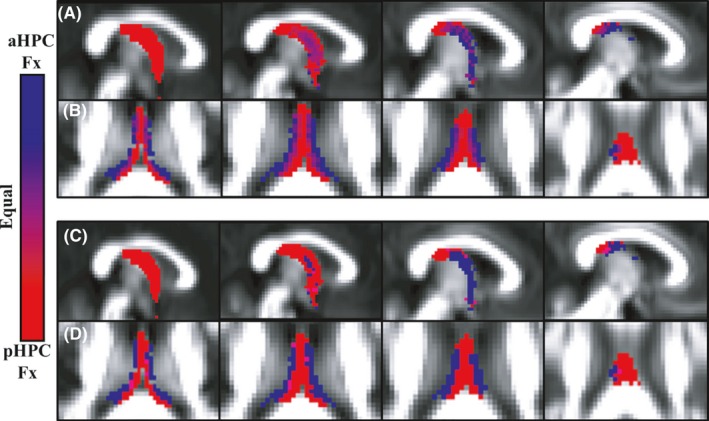
Figure 4.

(A, B) Relative probability maps depicting voxels that “belong” to the anterior (aHPCFx, blue) or posterior (pHPCFx, red) hippocampal fornix, with purple as equal ownership. The images in (A) go from the midline (left) to the lateral (right) parasagittal plane (MNI 2 mm y‐axis slices 45–48). The images in (B) proceed vertically from MNI z‐axis slice 44–47, that is, inferior (left) to superior (right). (C, D) Binary segmentations of anterior and posterior hippocampal fiber streams according to a winner‐takes‐all scheme (blue = anterior; red = posterior). Other conventions are the same as in (A and B)
