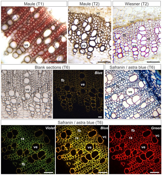
Figure 3.
Histochemical and epifluorescence tests in stems in secondary growth of Nicotiana tabacum. T1: Hand-sectioned fresh material submitted to the histochemical reactions. T2: Material fixed in Karnovsky’s solution, embedded in PEG, sectioned, and submitted to the histochemical reactions. T6: Material fixed in Karnovsky’s solution, embedded in PEG, sectioned, and stained. Violet, Blue, Green: Epifluorescence microscopy photographs were taken under violet, blue, or green light (indicated in each image). Scale bars: 50 µm. Abbreviations: PEG, Polyethylene glycol; fb, xylem fibers; ra, radial parenchyma; ve, vessel element; vc, vascular cambium.

