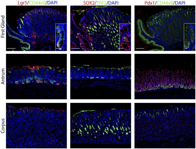
Figure 2.
The first gland possesses features similar to the deep antral glands. Each antibody labeling was imaged in first gland, antrum, and corpus sections. Left panels: In sections from Lgr5-GFP mice, immunofluorescence antibody labeling for Lgr5-positive progenitor cells using anti-GFP antibody (red) and anti-CD44v9 (green) and DAPI (blue). Middle panels: Immunofluorescence antibody labeling for transcription factor Sox2 (red) and TFF2 to label mucus neck cells (green) and DAPI (blue). Right panels: Immunofluorescence labeling of transcription factor Pdx1 (red) and CD44v9 (green) and DAPI (blue). All insets show enlarged view of the first gland. Scale bars for full image = 10 µm. Scale bars for insets = 3 µm. Abbreviations: GFP, green fluorescent protein; CD44v9, CD44 variant 9; DAPI, 4′,6-diamidino-2-phenylindole; TFF2, Trefoil Factor 2.

