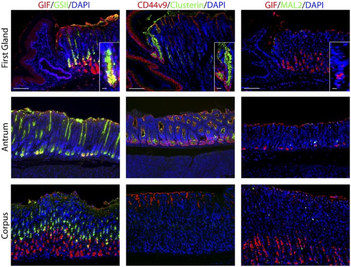
Figure 4.
Staining for putative metaplastic markers in the first gland. Each antibody labeling was imaged in first gland, antrum, and corpus sections. Left panels: Immunofluorescence antibody labeling using GIF (red) co-labeled with GSII-lectin (green) and DAPI (blue). Middle panels: Immunofluorescence antibody labeling for CD44v9 (red), Clusterin (green), and DAPI (blue). Note that staining of surface mucin with anti-CD44v9 is a consistent artifact. Right panels: Immunofluorescence antibody labeling for GIF (red) and MAL2 (green) and DAPI (blue). All insets show enlarged view of the first gland. Scale bars for full image = 10 µm. Scale bars for insets = 3 µm. Abbreviations: GIF, gastric intrinsic factor; GSII, Griffonia simplicifolia-II; DAPI, 4′,6-diamidino-2-phenylindole; CD44v9, CD44 variant 9.

