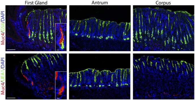
Figure 5.
The first gland cells express MUC4. Each antibody labeling was imaged in the first gland, antrum, and corpus sections. Upper panels: Immunofluorescence antibody labeling for MUC4 (red) co-labeled with GSII-lectin (green) and DAPI (blue). Lower panel: Immunofluorescence antibody labeling for MUC4 (red) co-labeled with UEA1-lectin to label surface cells (green) and DAPI (blue). Note that in addition to staining the first gland, MUC4 staining can also be observed in some endothelial cells in the corpus and antrum submucosa. All insets show enlarged view of the first gland. Scale bars for full image = 10 µm. Scale bars for insets = 3 µm. Abbreviations: GSII, Griffonia simplicifolia-II; DAPI, 4′,6-diamidino-2-phenylindole; UEA1, Ulex europaeus agglutinin-1.

