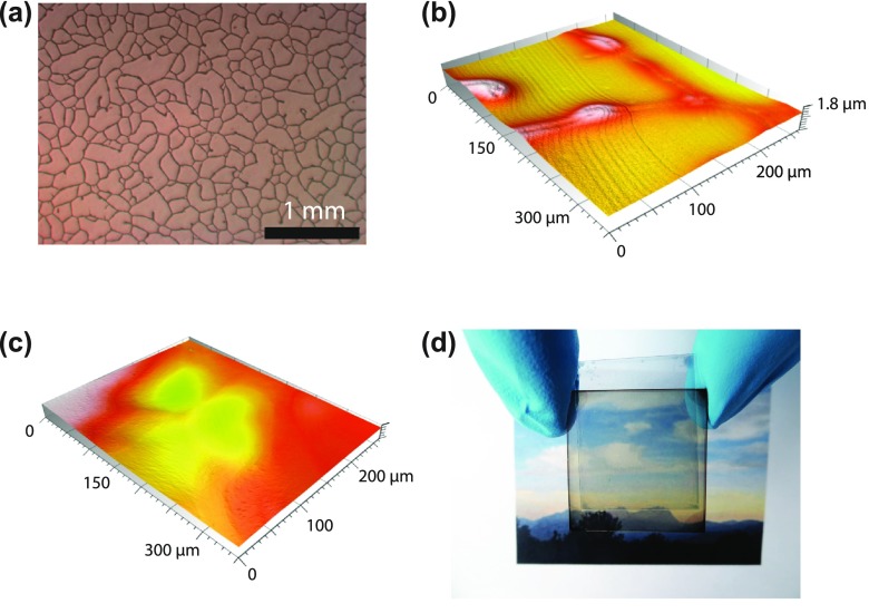
Figure 4.
(a) Optical microscopy image of the commercial substrate that consists of a random mesh-like silver network on PET. Optical confocal microscopy images of the laminate electrode when coating with a ~450 nm thick (b) and ~1.3 μm thick (c) PEDOT:PSS:sorbitol film. For small film thicknesses (b), the metal network is not fully covered. (d) Photograph of the semitransparent, laminated cell.

