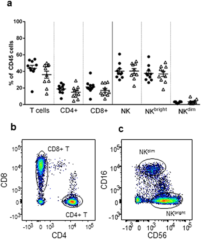
Figure 1. Phenotypic analysis of isolated CD45+ cells in the endometrium by flow cytometry.

Cells were isolated from the endometrium and stained with CD45-AlexaFluor-647, CD3-PECy5, CD4-FITC, CD8-PE, CD56-PECy7, CD16-APCCy7 antibodies. Endometrial immune cells from 10 control women (•) or 10 women with RM (Δ) were characterised and proportions of each cell population in the CD45+ lymphocyte gate displayed (a). Data displayed as mean ± S.E.M. Representative flow cytometry staining of endometrial CD4 and CD8 T cell populations in the CD45+/CD3+ cells (b) and NKbright (CD56bright/CD16−) and CD56dim (CD56+/CD16+) in CD45+/CD3− gating (c).
