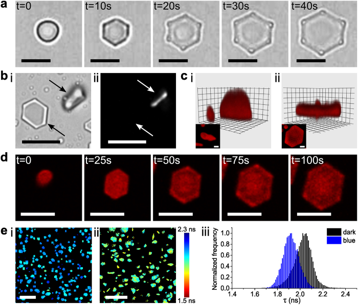
Figure 2.
(a) Time-lapse series of optical microscopy images showing blue light-induced transformation of a single trans-azoTAB:PAA irregular particle mounted on a highly PEGylated glass substrate into a trans/cis-azoTAB:PAA hexagonal platelet within 40 s; scale bars = 5 μm (see Supplementary Movie S1). Note the appearance of optically distinct regions at the corners of the hexagonal platelets after 20 s. (b) Optical (i) and polarized (ii) microscopy images showing two trans/cis-azoTAB:PAA hexagonal platelets oriented either face- or side-on (arrow) with respect to the substrate. Birefringence is only observed when the hexagonal axis is not perpendicular to the substrate; scale bars = 10 μm. (c) Confocal microscopy images of a single Nile red-stained trans-azoTAB:PAA microparticle (i) before and (ii) after in situ blue light irradiation showing formation of a trans/cis-azoTAB:PAA hexagonal platelet via lateral expansion and vertical contraction; insets show corresponding top views; scale bars = 1 μm. (d) Time-dependent video images showing extensive spreading of a single Cy5-ssDNA-containing trans-azoTAB:PAA microparticle mounted on a PEGylated glass substrate to produce a well-defined trans/cis-azoTAB:PAA hexagonal platelet; scale bars = 5 μm (see Supplementary Movie S2). (e) Fluorescence lifetime maps for sulforhodamine B-doped hydrated azoTAB:PAA microparticles mounted on a highly PEGylated glass substrate in the dark (i), or after blue light irradiation (ii); colours map different fluorescence lifetimes; scale bars = 10 μm. (iii) Corresponding fluorescence lifetime distribution histograms extracted from fitting of the FLIM images.

