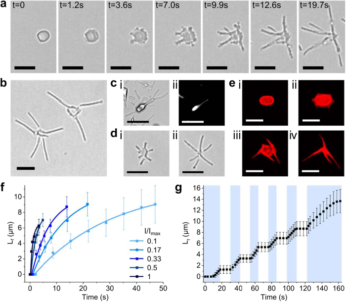
Figure 3.
(a) Time-lapse series of video images showing blue light-induced transformation of a single trans-azoTAB:PAA particle into a multipodal trans/cis-azoTAB:PAA micro-architecture within 20 s; scale bars = 5 μm. (b) Optical microscopy high magnification image showing two multipodal trans/cis-azoTAB:PAA micro-architectures comprising straight-sided filamentous outgrowths connected through a central core, scale bar = 5 μm. (c) Optical (i) and corresponding polarized (ii) microscopy images showing birefringence associated with the core and an adjacent filament of a multipodal trans/cis-azoTAB:PAA structure, scale bars = 5 μm. (d) Optical microscopy images showing simultaneous growth of straight-edged filaments from the corners of a single hexagonal platelet of trans/cis-azoTAB:PAA after exposure to blue light for (i) 10 s and (ii) 20 s to produce a discrete multipodal architecture; scale bars = 5 μm. (e) Time-lapse series of confocal fluorescence microscopy images of a single trans-azoTAB:PAA microparticle containing Cy5-ssDNA before (i) and after exposure to blue light for 10 s (ii), 45 s (iii) and 80 s (iv); scale bars = 5 μm. (f) Plots showing time-dependent increases in filament length (Lf) under blue light (turned on at t = 0) at varying light intensities (I/Imax). (g) Plot showing time-dependent changes in filament length (Lf) under repeated cycles of dark and blue-light irradiation (marked as white and blue regions, respectively).

