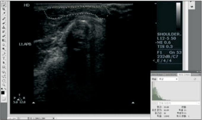
Fig. 3. Echo intensity (EI) analysis of ultrasound images. Grayscale images were used to determine muscle EI within each region of interest (ROI). The mean and standard deviation of the pixel brightness in each ROI were automatically calculated by Photoshop software.

