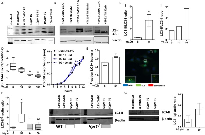
Figure 6.
Thioguanine (TG) promoted autophagy. TG administration enhanced autophagy in vitro: western blots for LC3-I, LC3-II and β-actin. (A) HeLa cells treated with dimethyl sulfoxide (DMSO) 0.1%,TG (10 or 100 µM)±Pepstatin A and E64D (PE) for 16 hours; (B) HT29, HCT116 and HepG2 cell lines treated with TG 50 µM for 16 hours; (C) LC3-II:LC3-I ratio in TG-treated (i) RAW cells and (ii) in HeLa+PE. Bacterial replication assay: (D) HeLa pretreated DMSO 0.1% or TG (10, 50 and 100 µM) infected with Salmonella (SL1344): luminescence readings 12 hours post-infection; SL1344 growth in media containing TG was not altered over that time period. N=4–8 experiments; (E) fluorescence microscopy quantification and representative image of LC3-II colocalisation with SL1344 bacteria-infected HeLa cells+TG 50 µM. Blue—4′,6-diamidino-2-phenylindole (DAPI) nuclear stain; red—Salmonella; green—LC3. Bars, mean±SD, N=3. Statistical analysis: unpaired t-test *p<0.05. The TG effect on autophagy was hypoxanthine (guanine) phosphoribosyltransferase (Hprt) dependent: (F) LC3-II to β-actin ratio quantification. Wild-type (WT) and Hprt−/−-derived primary murine fibroblasts treated with DMSO 0.1% or 50 µM TG for 16 hours. N=5–6. Western blot image. (G) WT colon organoids differentiated for 72 hours and then treated with DMSO 0.1% or TG 50 µM for 16 hours. Western blot image. LC3-II to β-actin ratio quantification, bars, mean±SD, N=2 from 6 wells/condition/replicate. Statistical analyses: Mann-Whitney non-parametric test: *p<0.05 WT TG 50 versus 0 µM; ##p<0.01 TG 50 µM Hprt−/− versus WT TG 50 µM.

