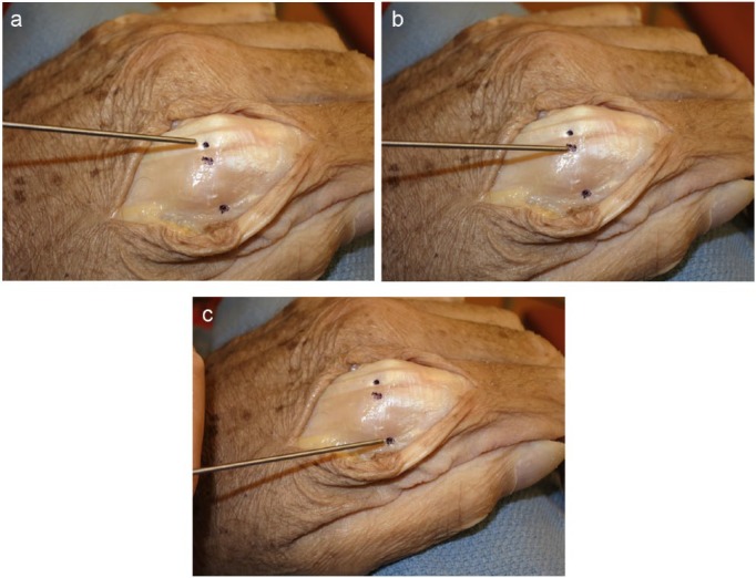
Figure 2.
Three different insertion points of the K-wires. (a) The first K-wire insertion was placed at a dorsal midline site, through the metacarpal head into the base of the P1, piercing the extensor tendon. (b) The second insertion site was through the metacarpal head adjacent to the extensor tendon. (c) The third K-wire was placed into the base of the P1 at the midaxial line, through the sagittal band, but remote from the fibers responsible for extending the PIP joint.
Note. PIP, proximal interphalangeal.

