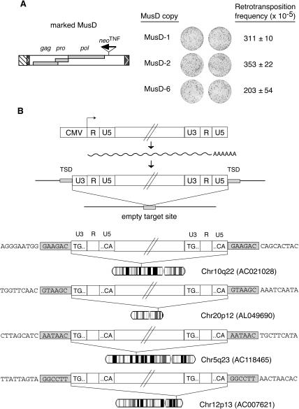
Figure 3.
Assay for autonomous MusD retrotransposition and characterization of the de novo insertions. (A) Each of the three MusD elements encoding functional proteins, as determined in the assay in trans, was marked with the neoTNF indicator downstream from the pol ORF as schematized and assayed for its autonomous retrotransposition. Images of the plates and retrotransposition frequencies are given as in Figure 2 (two to three independent experiments, with standard errors indicated). (B) Structure and chromosomal localization of four transposed MusD copies. The complete characterization of transposed MusD copies and insertion sites was performed using G418R clones from MusD-1 and MusD-6 marked elements assayed for transposition in HeLa cells. The structures of the marked MusD expression vector, of the intermediate MusD transcript, and of the de novo insertion (with reconstituted LTRs and target site duplication, TSD) are schematized, together with the corresponding empty target site. The sequences of four insertions are given below, with the [TG... CA] LTR borders and flanking DNA sequences indicated; target-site duplications (grayed) are found in all four cases, associated with complete LTRs. The GenBank Accession no. of each insertion site is given together with its chromosomal localization (with the R bands in white and the G bands in dark gray).

