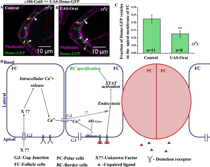
Fig 7. Elevation of Ca2+ stimulates vesicle internalization in the follicle cells.
(A-B): Maximum intensity projected images depicting the localization of Dome:GFP (Green) vesicles in the anterior follicle cells of indicated genotypes. Rhodamine Phalloidin staining (Magenta) marks the outlines of respective egg chambers. White arrowheads indicate vesicles localized to apical membrane of follicle cells. Yellow arrowheads mark cytoplasmic vesicles. (C): Histogram comparing apical fraction of Dome:GFP vesicles in the follicle cells for genotypes (A, B). (D): Proposed model of Inx2 mediated border cell fate specification. ‘n’ indicates number of egg chambers analyzed. Error bar represents Standard Error of Mean. ** represents p-value <0.01.

