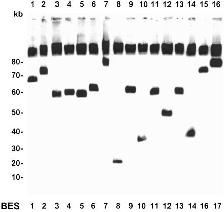
Figure 3.
Size determination of the T. brucei telomere clones. Genomic DNA of telomere clones was separated by FIGE gel electrophoresis. A representative telomere clone for the different ES types (only A sets) is shown, with the ES type indicated underneath. The gel was blotted and hybridized with vector pEB2 as probe. In addition to the telomere clone, this probe also hybridizes with yeast chromosomal DNA that is not separated under these conditions. Lanes are shown above, and size markers are indicated on the left in kilobases.

