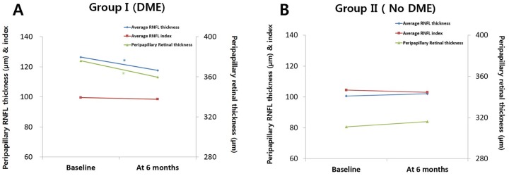
Fig 5. Average peripapillary retinal nerve fiber layer (RNFL) thickness, corresponding peripapillary retinal thickness, and RNFL index change during the 6-month follow up period in the 2 groups.
A) Eyes with diabetic macular edema (DME) show RNFL thinning (diamond marks) after anti-VEGF injection, but RNFL index after peripapillary retinal edema correction shows no change (rectangular marks). B) Eyes with no DME show no significant change in RNFL thickness (diamond marks) relative to the retinal thickness and RNFL index (rectangular marks) shows no change, similar to that in the DME group. *P < 0.01 (P values less than 0.01 were considered statistically significant).

