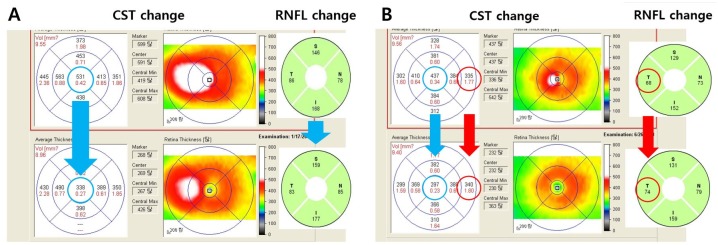
Fig 6. Spectral-Domain optical coherence tomography images (upper) before and (lower) 6 months after anti-VEGF injection for comparing changes between central subfield thickness (CST), temporal peripapillary retinal nerve fiber layer (RNFL) thickness in two patients with diabetic macular edema.
A) Case 1: The RNFL thicknesses in all quadrants decreased following the CST decrease. B) Case 2: Even though the central subfield thickness reduced (437 μm → 297 μm) after anti-VEGF injection, the temporal RNFL thickness increased (68 μm → 74 μm), as did the 6th subfield thickness (335 μm → 340 um).

