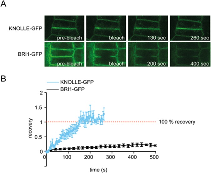
Fig 2. FRAP analysis of BRI1-GFP and KNOLLE-GFP in epidermal cells of Arabidopsis roots.
(A) Typical images of FRAP experiment prior and post bleach pulse of KNOLLE-GFP and BRI1-GFP. (B) Recovery-curves of KNOLLE-GFP (blue line) and BRI1-GFP (black line) in epidermal cells in the root meristem. Around 200 s after bleaching, fluorescence intensity of KNOLLE-GFP at the PM is fully restored. No such recovery is observed for BRI1-GFP. For KNOLLE-GFP n = 7, for BRI1-GFP n = 15, measured in independent replicas, error bars indicate standard error of means (SEM).

