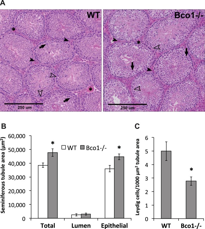
Fig. 3.
Histopathological and morphological assessment of testes reveals structural changes with Bco1 loss. A: pathological grading suggested increases in tubule size and moderate reductions in spermatocyte apoptosis in Bco1−/− mice, compared with WT. Representative images are shown; Leydig cells (solid arrowheads) reside in the interstitial space between seminiferous tubules, often alongside blood vessels (asterisks). Cells within the seminiferous tubules include Sertoli cells (open arrowheads) and spermatocytes (solid arrows). Morphological quantification was carried out as described in materials and methods and revealed that Bco1 disruption increased seminiferous tubule total and epithelial areas (B) and decreased Leydig cell number (C); n = 3–5 mice/genotype, *P ≤ 0.05.

