
Fig 6. The rate of amyloid plaque deposition was halted in APP mice whose slow oscillations were restored optogenetically.
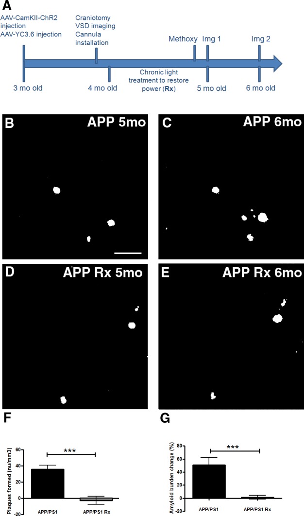
(A) Protocol for the experiment. Methoxy represents Methoxy-XO4 injection prior to imaging session 1 (Img 1). Methoxy was also injected prior to imaging session 2 (Img 2). (B-E) Representative in vivo multiphoton images show amyloid plaques within the same cortical field imaged in an APP mouse at 5 months (B) and 6 months of age (C). In vivo multiphoton images show amyloid plaques within the same cortical field imaged at 5 months (D) and 6 months of age (E) in an APP mouse whose slow oscillations were restored with light (Rx). (F,G) Bar graphs showing percentage change in amyloid plaque number (F) and amyloid plaque burden (G) between 5 and 6 months in APP mice or whose slow oscillations were restored with light activation of ChR2 (Rx) (n = 78–101 volumes in 7–9 mice/group). Scale bar, 100 μm. * p<0.05, *** p≤0.001.