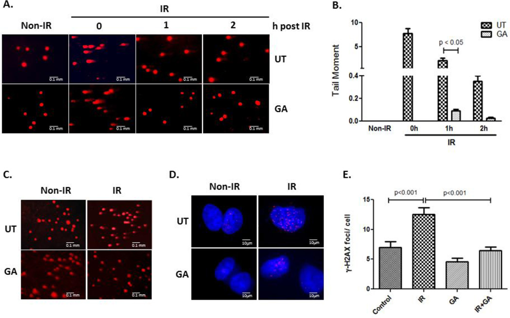
Figure 4. Resolution of radiation (IR)- induced DNA breaks in NS-SV-AC and shRNA TLK1 cells.
Single cells alkaline gel electrophoresis of A. NS-SV-AC cells and C. shRNA TLK1 cells. Cells were irradiated, and treated, or not, with GA. At indicated times, cells were electrophoresed, and cellular DNA analyzed by propidium iodide staining. B. The quantification of tail moment (mean ± SEM) of irradiated NS-SV-AC cells is graphed. D. Immuno-localization of phospho- S139 H2AX in NS-SV-AC cells 24 h after irradiation (IR). NS-SV-AC cells were irradiated prior to treatment with GA for 16 h. Cells were placed in drug-free medium for 8 h thereafter. Immuno-labeling with antiserum to gamma-H2AX (S139) is shown. E. Quantification of phospho-H2AX foci/ cell is shown. UT: untreated, IR: irradiation.

