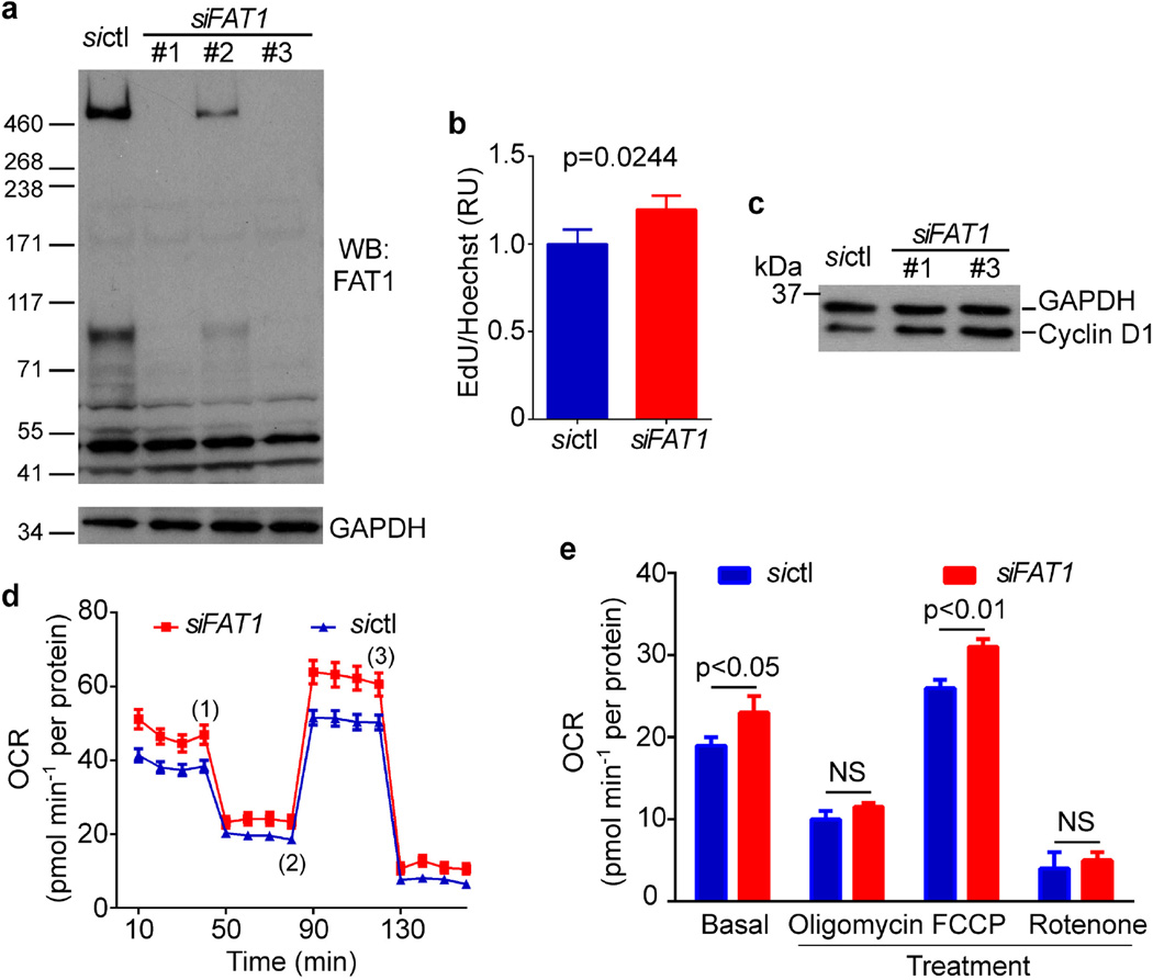
Extended Data Figure 8. FAT1 suppresses proliferation and mitochondrial respiration in human SMCs.
a, Western blotting for FAT1 expression in human aortic SMCs (HASMCs) treated with control siRNA (sictl) or FAT1 siRNAs (siFAT1) 1–3. For subsequent experiments, siFAT1 3 was used unless otherwise indicated. b, Proliferation of HASMCs after siFAT1 treatment, expressed as the ratio of EdU to Hoechst signal, normalized to sictl. n = 3, significance assessed by two-tailed t-test. c, Expression of cyclin D1 in sictl- or siFAT1-treated HASMCs. d, Oxygen consumption rate (OCR) of sictl- or siFAT1-treated HASMCs at baseline and in response to 2 µg ml−1 oligomycin (1), 3 µM FCCP (2), and 2 µM rotenone (3). e, Quantification of OCR from (d). n = 3, significance assessed by two-way ANOVA. All data shown as mean ± s.e.m. For gel source data, see Supplementary Fig. 1.

