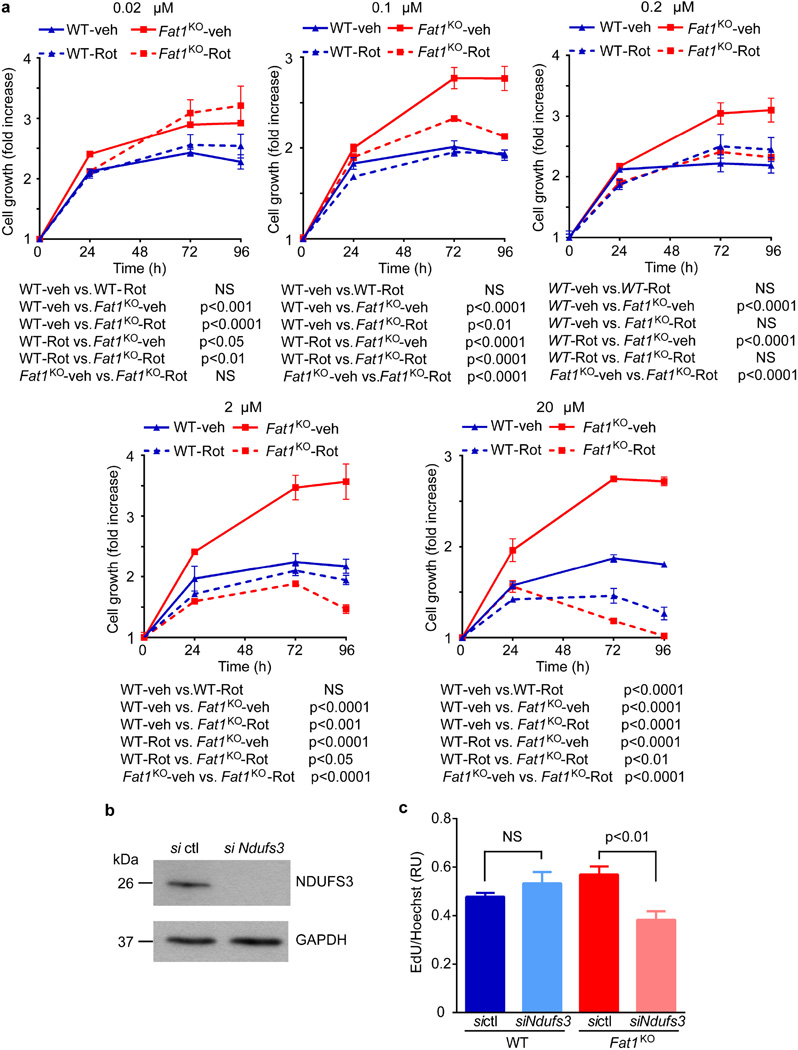
Extended Data Figure 3. Fat1 suppresses vascular SMC growth by inhibiting the electron transport chain.
a, Population growth of mouse aortic SMCs in the presence of various concentrations of rotenone, a complex I inhibitor. Addition of rotenone at concentrations from 0.1–2 µM did not compromise wild-type cell growth; by contrast, Fat1KO cell growth was suppressed to wild-type levels (n = 3); significance assessed by two-way ANOVA. b, Western blotting for NDUFS3 expression in mouse aortic SMCs treated with control siRNA (sictl) or Ndufs3 siRNA (siNdufs3). c, Proliferation of mouse aortic SMCs after siNdufs3 treatment, expressed as the ratio of EdU to Hoechst signal. RU, relative units. n = 3 for Fat1KO siNdufs3, n = 5 for other groups; significance assessed by one-way ANOVA. NS, not significant. All data shown as mean ± s.e.m. For gel source data, see Supplementary Fig. 1.

