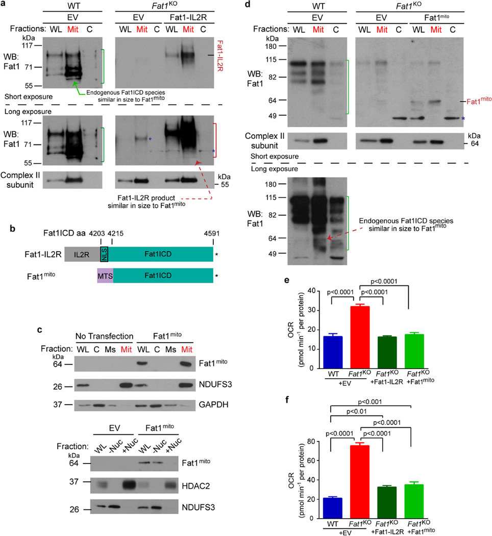
Extended Data Figure 5. Mitochondria-targeted Fat1 ICD is sufficient to repress oxygen consumption in vascular SMCs.
a, Electroporation of Fat1–IL-2R in mouse aortic SMCs, followed by subcellular fraction and SDS–PAGE analysis of Fat1 ICD fragments. Green bracket, endogenous Fat1 ICD species; red bracket, Fat1 ICD products from Fat1–IL-2R; blue asterisk, non-specific signal. b, Schematic of mitochondria-targeted Fat1 ICD, Fat1mito. Asterisk, stop codon. c, Top, Detection of Fat1mito in the mitochondrial fraction of Fat1mito -transfected 293T cells. Bottom, Exclusion of Fat1mito from the nuclear fraction of Fat1mito -transfected 293T cells. d, Electroporation of Fat1mito in mouse aortic SMCs, followed by subcellular fraction and SDS–PAGE analysis of Fat1 ICD fragments. Green bracket, endogenous Fat1 ICD species; blue asterisk, non-specific signal. e, f, Quantification of baseline (e) and maximal OCR (f) after introducing Fat1–IL-2R or Fat1mito into Fat1KO cells from Fig. 3a. Data shown as mean ± s.e.m., n = 15, significance assessed by one-way ANOVA. C, cytoplasmic; EV, empty vector; Mit, mitochondrial; Ms, microsomal; −Nuc, non-nuclear fraction; +Nuc, nuclear fraction; WL, whole-cell lysate. For gel source data, see Supplementary Fig. 1.

