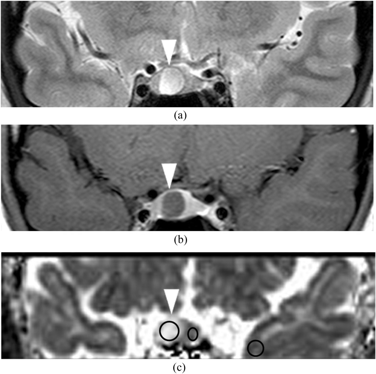
Figure 2.
A 23-year-old female with prolactin-producing microadenoma (0.28 cm3). T2 weighted image (a) shows a hyperintense mass (arrowhead) in the right side of the pituitary fossa. Post-contrast T1 weighted image (b) shows the hypointense mass (arrowhead). Apparent diffusion coefficient map (c) shows hyperintensity in the mass (2.54 × 10⁻³ mm2 s−1; arrowhead). Circles indicate the regions of interest in the adenoma, the unaffected anterior lobe of the pituitary gland and adjacent normal-appearing left temporal grey matter.

