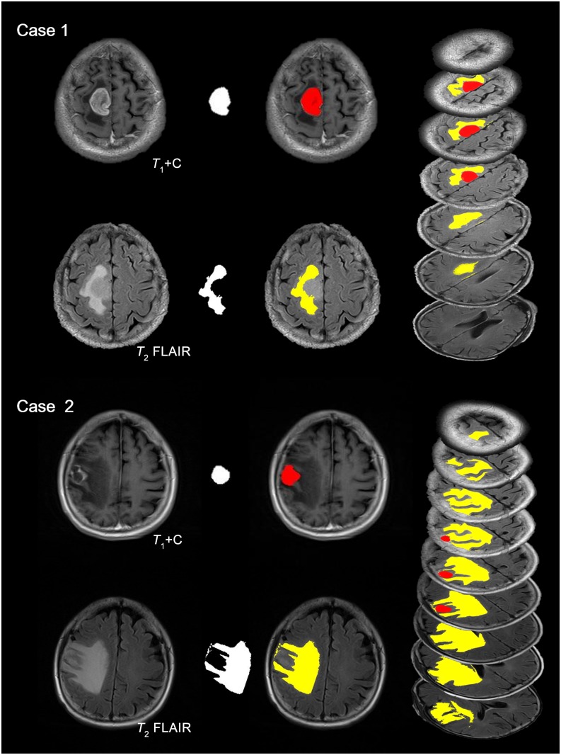
Figure 2.
Segmentation of tumour lesion and peritumoral oedema. Typical cases of glioblastoma multiforme (GBM) and brain metastasis are presented here. Case 1 is a patient with right frontal–parietal GBM (63 years old, male). Case 2 is a patient with single right frontal metastasis from lung cancer (60 years old, female). Enhanced T1 has been applied to identify the tumour lesion and T2 fluid-attenuated inversion recovery (FLAIR) for peritumoral oedema (first column). Segmentation results (second column) are enhanced with pseudocolour (third column). Tumour with necrosis is coloured red, while peritumoral oedema is in yellow. All slices are segmented and transformed to pseudo-three-dimensional images (fourth column). For colour image see online.

