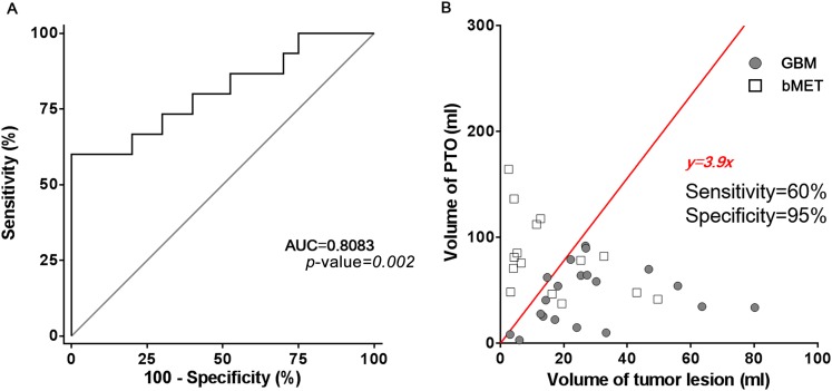
Figure 4.
Receiver-operating characteristic curve and linear regression of volume of peritumoral oedema (PTO) and lesion. (a) The receiver-operating characteristic curve shows the area under the curve (AUC) = 0.8083 with a significant difference (p-value = 0.002). (b) Linear regression shows that when PTO volume = 3.9 × tumour volume, glioblastoma multiforme (GBM) and brain metastasis (bMET) can be separated with a sensitivity of 60% and specificity of 95%. The ratio between volume of PTO and volume of tumour lesion can be a promising factor for separating GBM and bMET.

