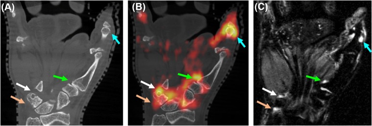
Figure 5.
Elevated fluorine-18 fludeoxyglucose (18F-FDG) uptake for active erosions in rheumatoid arthritis; co-registered coronal sections from (a) CT, (b) positron emission tomography (PET)/CT and (c) short tau inversion recovery MRI. The arrows indicate elevated 18F-FDG uptake sites of erosive changes (white, green and light blue arrows) and in the pisiform–triquetral compartment (light brown arrows). The linear range of the PET colour scale is 40–100% of the maximum standardized uptake value. For colour image see online.

